Welcome
A SMALL STEP

We take immense pleasure in introducing Bhargava eye centre, A complete eye centre situated in Bikaner (Rajasthan). Recognized as a premier eye care service provider,we have been rendering 18 Years of personalised eye care services with highly skilled doctors and fully trained staff who excels in soft skills.
From a humble beginning way back in year 1998, We have come a long way since to be recognized as one of the most trusted brands in eye care offering a complete spectrum of vision care treatments. We constantly strive to excel and are committed to serve every individual who walks through our doors.
This Hospital is fully equipped eye hospital with most advanced state of the art equipments in Ophthalmology.

Treatment Guide

Eye Care Tip

Wash the eyes with cold water in the mornings – it is soothing

more...
close
  • Wash the eyes with cold water in the mornings – it is soothing
  • Use sunglasses in sunlight to block the UV rays.
  • Always wear correct glasses & do not use someone else's glasses.
  • Do not read while lying down, in a moving train or bus.
  • Ensure there is proper light – do not read in dim light.
  • A well balanced diet is important. vitamin A which is an essential nutrient for eyes especially in children. Lack of vitamin A in children can cause night blindness.
  • If there is any family history of myopia, Glaucoma, Retinal detachment – periodic eye checkup is necessary.
  • If you have diabetes, hypertension (high blood pressure) periodic eye checkup is important.
  • Routine eye checkup for children is necessary to rule out possibility of eyeglass numbers.
  • If the child is squeezing his eyes while watching TV, rubbing eyes excessively or blinks too much, he might need glasses.
  • If the child has glasses number, it should be checked every six months to one year.
  • Do not let children play with sharply pointed objects like pencil, pen etc. many eyes are lost because of eye injury eg. bow & arrow.
  • In case of eye injury due to a fall or ball injury do not neglect it. The eye doctor should be consulted immediately.

Cataract

A cataract is an opacity or cloudiness in the natural lens of the eye.

more...
close

What is Cataract?
A cataract is an opacity or cloudiness in the natural lens of the eye. It is still the leading cause of blindness worldwide. In India it is commonly known as Safed Motia. When the lens is partially opaque, it is called an immature cataract and some light can pass through to help perform some routine functions. However, when the opacity increases to engulf the entire lens, vision is totally cut off and the cataract is mature.
What are the Problems Associated with Cataract?
Just as a smudged or dirty camera lens may spoil a photograph, opacity in the natural lens of the eye can result in a blurred image. Patients with cataracts usually complain of blurred vision either at distance, near, or both. This may interfere with tasks such as driving or reading. Other common complaints include glare, halos, and dimness of color vision. However, none of these symptoms are seen exclusively in cataract. Initially some help is achieved by changing the spectacle number, but in advance cases the spectacles also prove to be ineffective.
What are the causes of Cataract?
The development of cataracts in the adult is related to aging, sunlight exposure, smoking, poor nutrition, eye trauma, systemic diseases, and certain medications such as steroids.
What should one do?
The first thing a person must do on experiencing any of these symptoms is to consult an eye surgeon (Ophthalmologist) giving details of ones symptoms and getting ones eyes thoroughly examined. Answers to the following questions should be sought

  • Do I have cataract?
  • Do I have any other eye disease?
  • What is the cause of cataract in my eye?
  • What are the treatment modalities available?
  • What treatment would be most suitable for me?
  • What is the expected outcome of the treatment/surgery in my case?
  • What are the risks involved and possible complications?
  • How long can I wait before I get operated?
  • What does the surgery involve in terms of time and expenditure?
  • What is the treatment for Cataract?

There are no medicines to treat cataract. The answer lies only in surgery where the cataract is removed and replaced by an intra-ocular lens.
Where should I get my Cataract Operated?
The results of cataract surgery depend largely on skill of the operating doctor, the availability of latest equipment's and the technique of surgery and type of implant used. Eye Infections and damages (Surgically) are most difficult to treat hence reputed Centres where all facilities are Available and which have good patient inflow (Good Eye OPD) give best results. Specialized eye centres deal better in complicated cases
What is Phaco Technique?
Cataract surgery today is typically performed using a micro-incisional procedure called Phaco-emulsification. To the patient, this means minimal discomfort during or after surgery, a more speedy recovery of vision, and reduced risk of induced astigmatism. This means less dependence on glasses afterwards.
It is typically done through an incision less than 3 millimeters in size. Because of the careful construction of this incision, and its small size, the incision is generally self-sealing. This translates to a "no-stitch" type operation.
Phaco needle is introduced through the opening and ultrasonic vibrations are used to break the cataract into smaller fragments which are sucked out through this needle. An artificial lens called a foldable Intra-Ocular lens is now injected through this small opening. Once inside the eye, it opens up and serves to replace the original lens.
How is this Technique better than Conventional Technique?
The main advantages of Phaco are that a person has a good vision the very next day and may resume routine activities also the very next day. Stitches and related problems are things of the past. A person may now accomplish routine activities without spectacles, however a little spectacle correction may be required for fine focusing for distance and near. It is better for diabetic and hypertensive patients.
What to Expect After Surgery?
The great majority of patients may resume normal activities on the day of or day after surgery. Though the best vision may not be obtained until several weeks following surgery, but individual results vary considerably, depending on many variables. Activities such as reading, watching television, and light work will not hurt the operated eye.
Most surgeons arbitrarily recommend waiting 4 to 6 weeks before new glasses are prescribed. This allows the eye to achieve considerable stability from a refractive standpoint and, therefore, the glasses prescription should be accurate and relatively stable. Many patients are surprised at how clear their vision is after cataract surgery. Some patients may have better vision than they ever did before cataract surgery. Furthermore, depending on the degree of refractive error (need for glasses) prior to surgery, many patients will be much less dependent on glasses for far vision than they were before surgery. Patients will often notice that colors are brighter and more brilliant.
Is this Technique more Expensive than Conventional Stitch Technique?
Hi-tech expensive equipment and special lens other expensive material are required to execute a good surgery, making it a little more expensive than the conventional technique.
Can an Immature Cataract be Operated by Phaco?
It is easier and safer to operate on an immature cataract by Phaco. As the cataract matures, it tends to become harder requiring more Phaco energy to do the same job. Beyond a certain limit, excess energy may cause harm to the eye.
If one Eye has had a Conventional Cataract Surgery with IOL, can Phaco be done in the other Eye?
Yes.
What are the Type of Lenses Implanted After Cataract Surgery?
Foldable Lens: This lens has a diameter of 6.0 mm and is made of either Silicon or Soft Acrylic. On folding, it's diameter is reduced to 2.75 mm and it can be introduced into the eye through a 3 mm incision, where it unfolds automatically to take it's position. The main advantage of this lens is that there is fast visual recovery in the patient.
Non-foldable Lens: This lens has a diameter of 5.5 mm. A 3 mm incision has to be enlarged to 5.5 mm to introduce this lens. However, the incision still remains self-sealing and requires no sutures in most of the cases.
Aberration Free Foldable Lens: This lens is like a foldable lens in all ways, except that it's an aspheric lens. It reduces glare in the patient and is very useful for the patients who would like to drive at night.
Multifocal Lens: This is also a type of foldable lens which has distance as well as the near power in it. After it's implantation, patients will become much less dependent on glasses for near as well as distance. However, if you have cylindrical power in your glasses, you may need corrective lenses for fine work.
What Investigations are Required before Performing this Procedure?
The Blood Pressure (BP) and Blood Sugar should be in control.
E C G and a medical checkup may be required in some cases. Will I need to use Glasses After Phaco?
The lens that we use is a fixed powered lens. This is focused between the distance and the near vision more so for distance as this is the vision that is in use most of the time. As a result a person after the Phaco procedure will be able to perform routine activities without glasses. However a little spectacle correction would be required for fine focusing of distance and near vision.

Lasik & C-Lasik

LASIK (Laser assisted in-situ keratomileusis) is a refractive surgical procedure that

more...
close

LASIK (Laser assisted in-situ keratomileusis) is a refractive surgical procedure that results in rapid recovery of vision and has the capability to benefit patients with nearsightedness, farsightedness, and astigmatism by reducing dependence on eyeglasses and contact lenses.
Wave front Guided Lasik (Customized Lasik or C-Lasik) is fast emerging as a better tool to restore the quality of vision. The surgical procedure is similar in both the procedures and the common questions presented below are relevant to both.
How does the Wave Front Guided Lasik (C – Lasik) Work?
It is now known that sphere and cylinder are not the only vision disorders or aberrations in an optical system of a human eye. There is a second category of optical defects, called higher-order aberrations, that represent residual irregularities in the optical structure of the eye. Higher-order aberrations affect the quality of vision, The aberrometer which measures all the optical imperfection on a human eye. These higher order aberrations along with the usual sphere and cylinder aberrations can now be measured and documented with the help of a special machine called an aberrometer using the wave front technology Sophisticated software then converts the map into the clinical ablation profile prescription. The prescription is transferred electronically to the computer controlling the laser . The computer calculates the shot pattern (i.e., where and how the laser will operate on the cornea) required for the correction of both the patient's lower and higher order aberrations. These laser machines are the latest generation machines with special laser delivery systems capable of handling such corrections. Data map of optical imperfection in a human eye accessed by wave front technology which is then corrected by the laser
How is C – Lasik Betters than Simple Lasik Laser?
Lasik laser can correct the vision to normal (6/6 without glasses) in the absence of any other eye pathology. With C – Lasik, it is now possible in some cases to find a quality of vision, which in all be even better than what is achievable with simple Lasik laser.
What are the Minimum Requirments?
The person should be at least 18 years of age and should not have a significant increase in the prescribed power of spectacles in the past one year. Pregnant Women may not be suitable candidates.
How is Lasik Laser Performed?
The surgeon uses a microkeratome to create the flap. The flap is then positioned to one side of the corneal "bed" as the Excimer laser beam (left) is applied The laser ablation is completed and the flap is replaced. Once The flap is returned to preoperative position, the surgeon carefully checks and rechecks the flap to be certain it is secure and well-positioned. Lasik involves extensive computer analysis of the eye. Patient is then asked to come on the scheduled day for the laser treatment. Local anaesthetic eye drops are instilled in the eye to achieve a pain free procedure. The patient is now made to lie under the laser machine and asked to fix his gaze at a red light. The data of the patient is fed into the computer memory of the laser. A special machine called the microkeratome is used to pick up a thin corneal flap of 160 microns (equivalent to an onion skin).We use the best microkeratome that shapes the flap according to the cornea to give the best results. Laser is delivered to the cornea under this flap to correct the spectacle number. It takes 20 to 60 seconds to complete the laser delivery to one eye. The corneal flap is placed back in position where it holds strongly within two minutes due to its natural bonding properties. The entire procedure takes about 10 minutes.
Does the Procedure Hurt?
The laser procedure itself does not hurt. A slight pressure may be felt during the procedure. There is a possibility of some degree of discomfort after the procedure for 3 to 4 hours which is easily controlled by medication.
What to Except After Lasik Surgery?
our eye or eyes (if both are treated) will likely be quite blurry immediately after your LASIK surgery. Do not be alarmed. This is natural and expected. You will likely awaken the next morning after your LASIK surgery with much improved vision. Your vision should also improve over the first two to three weeks following surgery.
Will I be Totally Free From Spectacles?
yEs ,if you are a fit candidate ,lasik will give you unaided vision .In case of any difficulty you can be helped by your doctor
Are the Results Achieved From Lasik Permanent?
The effects of lasik on the cornea are permanent. Sometimes, internal changes within an eye may come with age and these may have some effect on the overall visual status. For example, a high myopia may progress even after the age of 18 years, or a person may require reading glasses after the age of forty like any normal individual
When can I Return to Work?
The results are often dramatic and very rapid, with most patients seeing well enough to drive a car without correction the very next day. However, the best post-operative visual acuity may not be obtained until 2 to 3 weeks, or in some cases, even a few months, after the procedure.
How Safe is this Procedure?
Lasik is a very safe procedure but like any other surgical procedure it can have some complications which are extremely rare if a proper screening check is done prior to the procedure. This includes measuring the corneal thickness with an ultrasound, mapping the corneal topography and a retina check. Nobody in our surgical experience has ever lost sight due to this procedure. This is US FDA approved procedure recommended for fighter pilots in USA.
What is the Worst Possible Complication?
Infection is a serious complication and could lead to deterioration of the vision. Infection is usually rare and can be well controlled with medications with little or no loss of quality of vision. At our centre, we have had no case of infection till date.
Can both Eyes be done the Same Day?
Yes, in most of the cases. However one eye may be done at a time by choice
How Long will I be on Medications?
Generally speaking a patient is on eye drops for 3 to 6 weeks. This is dependant of the level of correction attempted and the individual healing response.

ICL / C3R

The eye is like a camera in which lenses focus the picture on a light sensitive film.

more...
close

The Normal Cornea
The eye is like a camera in which lenses focus the picture on a light sensitive film. In the human eye, the transparent cornea and lens focus light on the retina, which changes it into electrical signals, which are then transmitted to the brain by the optic nerve to be perceived as images. The cornea is the front transparent window of the eye and forms the outermost one-sixth of the eyeball. It is lamellar in nature (like plywood) and is made up of 5 layers, each of which has a definite function. In order to be effective it must remain transparent. Freezing, heating, molding, lathing, tattooing, excising, incising and transplanting are all means by which the delicate and sensitive cornea has been altered for optical, therapeutic and cosmetic purposes. Due to absence of blood vessels in the cornea, much of its oxygen requirement comes from atmospheric oxygen dissolved in the tear film. When the eyelids are closed, oxygen enters the cornea from the superficial conjunctival vessels. Nutrients needed for the cornea pass into it by diffusion. Hence, carbon dioxide and waste products are also removed across the tear film. Hence, any deficiency of the tear film will directly or indirectly affect the cornea.
What is Keratoconus?
Normally the cornea is nearly spherically shaped thus allowing light to be focused clearly on the back of the eye (retina). However in a condition called Keratoconus, the cornea begins to thin, and this allows the normal pressure of the eye to make the cornea bulge forward taking on a cone-shape. As the cornea gradually becomes more cone-shaped, the vision blurs and becomes distorted due to a high degree of astigmatism. Initially vision may be correctable with spectacles, but as the condition progresses, and the cornea becomes more irregular causing distorted vision, spectacles become less effective. In such a situation, contact lenses not only provide better vision, but also help to retard the progress of the disorder. A rigid contact lens (RGP / "semi-soft" contact lenses) must be used, so that it can hold its shape, as a soft lens would simply mould to the existing shape and thus not allow complete correction of the problem. Sometimes the patient is fitted with soft lenses (for comfort), over which semi-soft lenses are fitted ("piggy-back" lenses).
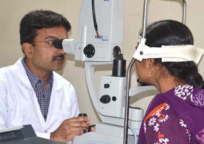
Fitting contact lenses for keratoconus requires expertise. Well-fitting contact lenses dramatically improves such a patient's vision to nearly that of a normal person's, and significantly improves his or her quality of life. Any excessive pressure of a poorly fitting lens on the cone apex can cause permanent scarring within months or years (This scarring can also occur naturally). For this reason it is important for regular follow-up visits to be made so that any corneal changes that have occurred can be compensated for in the design of a new lens. It is quite common for patients to be refitted at irregular intervals as the condition progresses. Rarely, scarring is so severe that a corneal graft (transplant) is necessary.
A recent promising treatment modality for keratoconus is C3R (Corneal Collagen Cross-linking).
What is Corneal Collagen Cross-linking (C3R)?
Cross Linking of Cornea Collagen (C3R) is a process to increase the mechanical stability of corneal tissue. The aim of this treatment is to create additional chemical bonds inside the corneal stroma by means of a highly localized photo polymerization. The indications for cross linking today are corneal ectasia the disorders such as keratoconus and pellucid marginal degeneration, iatrogenic keratectasia after refractive lamellar surgery and corneal melting that is not responding to conventional therapy.
The History of Corneal Cross-linking
The procedure was developed from 1993 till 1997 by Prof. Theo seiler and Prof. Eberhard Spoeri at the University of Dresden, Germany. The first patients were treated in 1998. Today corneal cross-linking is performed in more than 300 centers around the world. Corneal Cross-linking has the potential to become the standard treatment for keratoconus thus preventing the need for penetrating keratoplasty!
The Principle of C3R
Photo-polymerization using UV-light was found to be the most promising technique to achieve cross-links in connective tissue. Photo-polymerization is activated by means of a non-toxic and soluble photomediator and a wavelength which is absorbed strongly enough to protect deeper layers of the eye (riboflavin-UVA technique).
UV-A radiation with concomitant administration of riboflavin solution leads to physical cross linking of the corneal collagen fibers.
Thus progressive corneal thinning is slowed down or even stopped and the Bio-mechanical strength of corneal tissue is improved.
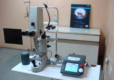
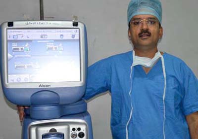
The Device for C3R
For C3R we need riboflavin dye and a special device called cross-linker. Cross linker is a device to deliver UV-A light of specific wavelength of 365 nm , at controlled energy level of 3 mW/cm.sq.
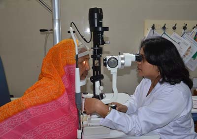
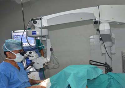
The C3R Procedure
After removal of the corneal epithelium, riboflavin solution is instilled for 30 minutes on to the cornea. Then the corneal penetration of this is checked by establishing that the anterior chamber is slightly yellow. Pachymetry is performed to make sure that minimum corneal thickness is maintained.
UV-A radiation starts under continued administration of Riboflavin Solution. After 30 minutes of radiation treatment is finished and the patient receives post-operative treatment like after a PRK procedure. A bandage contact lens is inserted in the operated eye and the patient is administered oral and topical antibiotics, steroids, anti-inflammatory medication as well as lubricant eye drops.
Clinical Experience with C3R
Today, more than 1,400 eyes have been cross-linked world wide in controlled clinical studies with a follow up to 5 years.
Clinical studies have shown a significant increase in best corrected visual acuity (BCVA) in more than 85% of the treated eyes.
Six months after corneal cross-linking the refractive cylinder was reduced in over 80% of the eyes. The steepest K-value was usually decreased by 1 diopter and the percentage of eyes that had a clinical relevant reduction exceeds 86%.
Safety of C3R
Corneal cross-linking is considered to be a safe procedure, provided the recommended safeguards are observed. Up until today no sight threatening side effects have been reported.
An excellent Vision Correction Option
The ICL (Implantable Contact Lens) is a state-of-the-art refractive error solution that is ideal for anyone who has the need or desire for removal of power with high quality of vision correction. ICL or Implantable Contact Lens, as the name suggests, is a kind of lens which is implanted into the eye and does not require frequent removal like a normal contact lens. This phakic intraocular lens has numerous advantages including its correction of the widest range of myopia (near sightedness), hyperopia (far sightedness) and astigmatism (cylindrical power). * ICL can correct a wide range of vision errors by permanently inserting a Contact lens in front of the natural lens of the eye.

  • ICL is a kind of soft contact lens which is inserted into the eye through a very small incision
  • Just like LASIK or Wavefront Lasik it takes only 5-10 minutes for the procedure
  • The lens is customized according to each eye's shape and size
  • Widest power correction range from +10D to -20D with cylinder upto 6D
  • Made from a material called "Collamer" which is bio compatible (safe to stay in the eye for very long time)
  • This new technique is similar to cataract surgery, but the natural lens remains in place so the eye's natural focusing ability is preserved.
  • An implantable contact lens is beneficial because it becomes a permanent fixture of the eye, avoiding time consuming maintenance.
  • It does not get lost, or have to be replaced like glasses and contact lenses.
  • ICL procedures are being used on highly nearsighted and farsighted patients who may not be candidates for the more common laser procedures such as LASIK, LASEK, and PRK. Unlike laser vision correction procedures that permanently change your vision, it is possible to later remove an ICL.

How do I know if I am a suitable ICL candidate?

  • Candidates for the ICL are above 18 years of age, suffer from myopia (nearsightedness),hyperopia (farsightedness) and/or astigmatism (cylindrical power) and want to experience superior vision correction.
  • Candidate with refractive error who are unsuitable for laser refractive surgery.
  • Prospective person should consult his/her ophthalmologist (eye surgeon) for more information, including an assessment of their candidacy.
  • Women who are pregnant or nursing should wait to have the ICL implanted. Lastly, those without a large enough anterior chamber depth or endothelial cell density may not be a good ICL candidate.

Advantages of ICL?

  • No blood! No pain! No hospitalisation!
  • Almost all levels of power can be treated
  • Excellent quality of vision
  • Easily removed or replaced (5-10 minutes)
  • Cosmetically good as it's INVISIBLE!!
  • Fast recovery

Why Patients Seek ICL
Patients seek the ICL because they expect the highest quality of results from their refractive vision correction procedure. The advantages of this phakic IOL (highlighted below) make the ICL a sought after treatment for nearsightedness & farsightedness.

  • High quality of vision The ICL not only corrects your refractive power or number, but it also enhances your quality of vision by producing sharp vision.
  • Wide treatment range In comparison to other refractive procedures, the ICL offers the widest treatment range for correction of vision.
  • Foldable because the ICL is foldable, a small incision is required during the procedure. This feature makes the procedure efficient (no sutures needed) and improves healing time.
  • Invisible the placement of the ICL into the posterior chamber of the eye makes the lens invisible to both the patient and any observer.
  • Collamer composition Collamer is made from collagen, which is a substance that naturally occurs in the body. This makes the lens highly biocompatible with the eye.
  • Proven track record Implanted in over 65,000 eyes worldwide, the safety and amazing improvement in vision quality of the ICL has been proven over the last 15 years. Because the ICL is also ideal for patients with contraindications for laser refractive surgery, some people may consider the ICL as an alternative to LASIK; however, as you can see, it is so much more than that. It is often compared to the corneal refractive procedure because the ICL takes refractive surgery beyond the limits of LASIK. Patients, who may not discover the ICL until they begin to search for an alternative treatment to avoid LASIK or PRK, realize that the ICL is not just an alternative; it is the good choice for superior vision quality.
  • Advantages of ICL

    • Preserves accommodation
    • No corneal tissue removed
    • Retains corneal asphericity
    • Possibly retains contrast sensitivity
    • Removable

    How does the ICL work?

    • Similar to a contact lens
    • Designed to remain inside the eye
    • Doesn't get dirty and needs no maintenance unlike a contact lens
    • Once-a-year visit to hospital recommended for examination

    How does the ICL differ from other refractive procedures?

    • Does not cut or remove tissue from the cornea
    • Cornea retains it natural shape
    • Safer for higher degrees of myopia, hyperopia, astigmatism and thin corneas
    • Less glare issues on patients with large pupils
    • Very stable over time, no regression

    What to expect on the procedure?

    • Procedure should take 10-15 minutes per eye
    • Laser Iridotomies done prior to surgery
    • Dilating and anesthetic drops

    The ICL Procedure:

    • The implant surgery is quick and painless, lasting only about 10 – 15 minutes.
    • The area around your eyes will be cleaned and a sterile drape may be applied around your eye.
    • Eye drops or a local anesthetic will be used to numb your eyes.
    • When your eye is completely numb, an eyelid speculum will be placed between our eyelids to keep you from blinking during the procedure.
    • The recovery time is short and the results of the surgery are almost immediate.
    • Most patients resume normal activities within a week.

    Potential ICL risks include:

    • Overcorrection: This complication occurs if the prescriptive power of the implanted ICL is too strong. In most cases it can be corrected with corrective eyewear or with an ICL replacement.
    • Undercorrection : The opposite of overcorrection, undercorrection is the result of an implantable contact lens with too weak of a prescription.

    Correction methods are similar to those of overcorrection.

    • Infection During most surgeries, there is a potential of an infection.
    • Increased intraocular pressure Pressure may build in the eye after an ICL procedure. The sooner a surgeon is alerted to this, the greater the chance of avoiding serious damage.
    • This is detected during your follow up visits with us or in case you face acute blurring of vision or headaches, you must visit the eye clinic
    • ICLs have the potential, however slight, of needing to be repositioned.
    • Damage to crystalline lens : because implantable contact lenses are implanted into the eye, there is a potential that the eye's natural lens may be damaged during the procedure. If the damage is severe, the crystalline lens may need to be replaced with an intraocular lens.
    • Cataract development : Over 50 percent of the population will develop cataracts by the age of 65, however, it is believed that the use of some implantable contact lenses may cause cataracts at an earlier age, this however is rare.
    • Halos, glare, and double vision : Updated ICL models greatly diminish the risks of halos, glare, and double vision.
    • Retinal detachment : Less than 1 percent of patients in the clinical studies were affected by retinal detachment. It should be noted, however, that the occurrence of retinal detachment increased as the degree of myopia increased.

    Where is the ICL placed?
    A trained ophthalmologist will insert the ICL through a small micro-incison, placing it inside the eye just behind the iris in front of the eye's natural lens. The ICL is designed not to touch any internal eye structures and stay in place with no special care.

    What is Toric ICL?
    The Toric ICL is only a variant of ICL. Toric ICL corrects your nearsightedness as well as your astigmatism (cylindrical power) in one single procedure. Each lens is custom made to meet the needs of each individual eye.

    What is the track record of the ICL?
    Prior to being placed on the market, the ICL was subject to extensive research and development. Today,more than 65,000 patients worldwide enjoy the benefits of the device. In an USFDA clinical trial, over 99 percent of patients were satisfied with their implant. The ICL has a track record of stable, consistently excellent clinical outcomes. The lens has been available internationally for over 12 years.

    Does it hurt?
    No, most patients state that they are very comfortable throughout the procedure. Your ophthalmologist will use a topical anesthetic drop prior to the procedure and may choose to administer a light sedative as well.

    What is the ICL made of?
    The ICL is made of Collamer®, a highly biocompatible advanced lens material which contains a small amount of purified collagen. Collamer does not cause a reaction inside the eye and it contains an ultraviolet filter that provides protection to the eye. Collamer is a material proprietary to STAAR Surgical Company, the company that manufactures ICL.

    What if my vision changes after I receive the ICL?
    One advantage of the ICL is that it offers treatment flexibility. If your vision changes dramatically after receiving the implant, your doctor can remove and replace it. Patients can wear glasses or contact lenses as needed following treatment with the ICL. The implant does not treat presbyopia (difficulty with reading in people 40 and older), but you can use reading glasses as needed after receiving the ICL.

    What type of procedure is involved in implanting the ICL?
    The implantation procedure for the ICL (Implantable Contact Lens) is refractive eye surgery that involves a procedure similar to the intraocular lens (IOL) implantation performed during cataract surgery. The main difference is that, unlike cataract surgery, the ICL eye surgery does not require the removal of the eye's natural lens. The ICL procedure is a relatively short outpatient procedure that involves several important steps. The surgical procedure to implant the ICL is simple and nearly painless.
    As a ICL candidate, your doctor will prepare your eyes one to two weeks prior to surgery by using a laser to create a small opening between the lens and the front chamber of your eye (iridotomy). This allows fluid to pass between the two areas, thereby avoiding the buildup of intraocular pressure following the surgery. However, some surgeons choose to do this step on the same day of the surgery. The implantation procedure itself takes about 10-15 minutes and is performed on an outpatient basis, though you will have to make arrangements for someone to drive you to and from the procedure.
    You can expect to experience very little discomfort during the ICL implantation. You will undergo treatment while under a light topical or local anesthetics. Following surgery, you may use prescription eye drops or oral medication. The day after surgery, you will return to your doctor for a follow-up visit. You will also have follow-up visits one month and six months following the procedure. Although the ICL requires no special maintenance, you are encouraged to visit your eye doctor annually for check-ups following the ICL procedure.

    Can the ICL be removed from my eye?
    Although the ICL is intended to remain in place permanently, a certified ophthalmologist can remove the implant in a very quick & short procedure.

    Is the ICL visible to others?
    No, the ICL is positioned behind the iris (the colored part of the eye), where it is invisible to both you and observers. Only your doctor will be able to tell that vision correction has taken place.

    Will I be able to feel the ICL once it is in place?
    The ICL is designed to be completely unobtrusive after it is put in place. It stays in position by itself and does not interact with any of the eye's structures.

    Where can I get my ICL procedure done?
    Please be aware that ICL procedure is presently available at select centre`s & hospitals in India as it requires precision and skills. In our Institution our Cornea and LASIK experts will guide you better if you are the right candidate for this procedure.

    Glaucoma

    Glaucoma is the second leading cause of blindness in the world

    more...
    close

    Glaucoma is the second leading cause of blindness in the world, according to the World Health Organization. Estimates put the total number of suspected cases of glaucoma at around 65 million worldwide.
    It is a group of eye diseases that gradually steal sight without warning. In the early stages of the disease, there may be no symptoms. Experts estimate that half of the people affected by glaucoma may not know they have it. Four Key Facts About Glaucoma
    Glaucoma is a leading cause of blindness
    There is no cure (yet) for glaucoma
    Everyone is at risk for glaucoma
    There may be no symptoms to warn you
    Vision loss is caused by damage to the optic nerve. This nerve acts like an electric cable with over a million wires. It is responsible for carrying images from the eye to the brain. There is no cure for glaucoma-yet. However, medication or surgery can slow or prevent further vision loss. The appropriate treatment depends upon the type of glaucoma among other factors. Early detection is vital to stopping the progress of the disease. It was once thought that high pressure within the eye, also known as intraocular pressure or IOP, is the main cause of this optic nerve damage. Although IOP is clearly a risk factor, we now know that other factors must also be involved because even people with "normal" levels of pressure can experience vision loss from glaucoma.
    What are the common types of glaucoma?
    Adult glaucoma falls into two categories- open angle glaucoma and closed angle glaucoma . These are marked by an increase of intraocular pressure (IOP), or pressure inside the eye. When optic nerve damage has occurred despite a normal IOP, this is called normal tension glaucoma. Secondary glaucoma refers to any case in which another disease causes or contributes to increased eye pressure, resulting in optic nerve Symptoms of angle closure glaucoma may include headaches, eye pain, nausea, rainbows around lights at night, and very blurred vision.
    Are You at Risk For Glaucoma?
    Everyone is at risk for glaucoma. However, certain groups are at higher risk than others. People at high risk for glaucoma should get a complete eye exam, including eye dilation, every one or two years
    The following are groups at higher risk for developing glaucoma
    People Over 60
    Family Members with Glaucoma
    Steroid Users
    Eye Injury
    Other Risk Factors
    High Myopia (nearsightedness)
    Diabetes
    Hypertension
    Central corneal thickness less than .5 mm
    What is involved in a glaucoma evaluation?
    Our specialists continuously fight the severe consequences of not detecting the disease in its early stages To achieve an accurate assessment, experienced ophthalmologists perform a comprehensive glaucoma screening that consists of three non-invasive, pain-free procedures:

    • Measurement of intraocular eye pressure (IOP) with Applanation tonometry.
    • Gonioscopy -To assess type of glaucoma and management options
    • Assessment of the optic nerve .A non-invasive slit lamp biomicroscope is used to determine whether or not there are changes in the optic nerve in the diagnosis for glaucoma.
    • Assessment of retinal nerve fiber layer It is earliest site of damage. We have GDx VCC (Carl zeiss ,Germany) for earliest glaucoma detection.
    • Evaluation of a patient's visual field with Humphreys Perimeter, Zeiss Germany Glaucomatous damage produces characteristic defects in the visual field.

    Glaucoma treatment seeks to decrease intraocular pressure and prevent damage to the optic nerve. Different types of glaucoma require different therapies to prevent further damage to the eye's structures. At the beginning of treatment, the doctor will generally recommend medication or a combination of medications for the specific condition.


    Therapies may include:
    Eye drops (or a combination of eye drops and pills) to reduce intraocular pressure. Several different classes of glaucoma medications are available to provide pressure reduction including beta blockers, prosaglandin analogues, alpha adrenergic agaonists, miotic, epinephrine compounds, and oral and topical carbonic anhydrase inhibitors. These medications work by either reducing the rate at which fluid in the eye is produced or increase the outflow of fluid from the eye.
    Laser treatment to open the drainage angle and reduce intraocular pressure.
    Surgery to create a new passage for fluid drainage. Surgery is usually reserved for cases that cannot be controlled by medication and following appropriate laser treatment.
    Is blindness due to glaucoma preventable?
    Regular diagnostic examinations by an ophthalmologist are the key to preventing loss of vision due to glaucoma. Ophthalmologists are medical doctors, specialists in eye care and trained to examine and treat eye diseases. Although there is no way to reverse damage, if glaucoma is diagnosed and treated early, blindness almost always is preventable.
    What are the symptoms of glaucoma?
    Although the blindness associated with this disease is preventable, more than one million people in the United States have some glaucoma-related vision loss. In most cases, glaucoma is asymptomatic (has no symptoms). By the time an individual experiences decreased vision, the disease is frequently in its latter stages. Since early warning signs of glaucoma are rare, it is important --- especially for those at risk --- to have medical eye examinations at appropriate intervals, as described in this section.
    Symptoms depend on the type of glaucoma the individual has Those who have chronic glaucoma may not be aware of any symptoms because the disease develops slowly and patients rarely notice loss of peripheral vision. Those who have an acute form of glaucoma may develop severe symptoms because ocular pressure rises quickly and they may experience:

    • Blurred vision, especially at night
    • Halos or rainbows around lights
    • Severe headaches or eye pain
    • Nausea

    How does glaucoma affect the eye?
    The eye has an internal pressure created by production of a clear fluid called aqueous humor. This fluid circulates through the eye and exits through the anterior chamber angle and ultimately drains into the blood stream. In glaucoma, the aqueous humor outflow is obstructed, resulting in increased eye pressure and, eventually, optic nerve damage.

    Eyedrop Tips
    Prescription eye drops for glaucoma help maintain the pressure in your eye at a healthy level and are an important part of the treatment routine for many people. Always check with your doctor if you are having difficulty. Remember
    Follow your doctor's orders. Be sure your doctor knows about any other drugs you may be taking (including over-the-counter items like vitamins, aspirin, and herbal supplements) and about any allergies you may have.
    Wash your hands before putting in your eye drops.
    Be careful not to let the tip of the dropper touch any part of your eye.
    Make sure the dropper stays clean.
    If you are putting in more than one drop or more than one type of eye drop, wait five minutes before putting the next drop in. This will keep the first drop from being washed out by the second before it has had time to work. Store eye drops and all medicines out of the reach of children. Steps For Putting In Eye Drops

    • Start by tilting your head backward while sitting, standing, or lying down. With your index finger placed on the soft spot just below the lower lid, gently pull down to form a pocket.
    • Let a drop fall into the pocket.
    • Slowly let go of the lower lid. Close your eyes but try not to shut them tight or squint. This may push the drops out of your eye.
    • Gently press on the inside corner of your closed eyes with your index finger and thumb for two to three minutes. This will help keep any drops from getting into your system and keep them in your eye, where they are needed.
    • Blot around your eyes to remove any excess. If you are still having trouble putting eye drops in, here are some tips that may help.

    Retina

    Diabetes affects the retina and this condition is called as diabetic retinopathy.

    more...
    close

    How does myopia affect the retina?
    Diabetes affects the retina and this condition is called as diabetic retinopathy. It is a micro angiopathy affecting the retinal blood vessels. The main features of diabetic retinopathy are micro vascular (small blood vessel) occlusion and leakage. As a result of this, there is reduced blood supply to the retina .Due to leaking blood vessels, hemorrhages and fluid accumulation in the retina can occur.
    This initial stage is also called as Non-Proliferative Diabetic Retinopathy (NPDR).
    What happens in late stages of Diabetic Retinopathy?
    As a result of reduced blood supply to the eye because of diabetes the eye will start forming its own new blood vessels Proliferative Diabetic Retinopathy (PDR), which are fragile hence they can rupture and bleed any time which lead to sudden drop in vision. This is known as Vitreous hemorrhage. Sometimes in more severe forms traction on retina can develop that can lead to retinal detachment. This requires treatment in the form of laser to the eye or surgery or both. The treatment is to stabilize the patient's vision and prevent further progression of visual loss. Proliferative Diabetic Retinopathy
    What is Diabetic maculopathy?
    Diabetic maculopathy is the commonest cause of visual loss in diabetic patients. Fluid accumulation leads to swelling in the macula which is the area concerned with vision. It may be focal, diffuse or ischemic.
    Focal-focal leakage from the microaneurysms which leads to localized swelling.
    Diffuse-diffuse leakage which leads to swelling throughout the posterior pole.
    Ischemic-in this type the blood supply to the macula decreases and this leads to poor vision.
    Diabetic Maculopathy
    Lasered Ratinopathy
    What are the different investigations done in Diabetic Retinopathy?
    Fundus Fluorescein Angiography (FFA) and Optical Coherence Tomography (OCT) are the two investigative procedures which may be done before the treatment plan for Diabetic Retinopathy. Fundus Fluorescein Angiography (FFA)
    This is a diagnostic procedure where in fluorescein dye is injected into a vein of the hand and then a series of photographs of the patient's retina are taken. This gives us a picture of the passage of the dye through the microvasculature of the eye. This helps us to know the status of the blood vessels in retina and reveals blockage, excess leakage of the dye in case of abnormal new blood vessels. It also helps us to know whether the patient needs laser photocoagulation or not.
    What is the treatment of the Diabetic Retinopathy?
    The treatment is usually in the form of laser or rarely an injection is given in the eye.
    The laser treatment may be done in 1- 3 sittings
    How frequent should a diabetic have eye check up?
    Every patient once diagnosed to have diabetes should have regular eye check ups every 6 months.
    What are the parts of retina?
    Macula (central retina) – appreciation of fine details, reading and color vision
    Peripheral retina – functions for night vision
    What are the symptoms of retinal disease?
    Sudden or gradual decrease in vision
    Loss of field of vision
    Flashes of light
    Floaters
    Objects appearing distorted (Metamorphopsia)
    Night Blindness
    When should one consult a retinologist?
    Whenever patient has any of the above mentioned symptoms he is advised to have an immediate check up with a retina specialist. Regular eye examinations if the patient is diabetic, hypertensive and over forty years.
    What are the diseases that affect the retina?
    Common diseases include
    Diabetes mellitus
    Hypertension
    Age related macular degeneration
    Myopia
    Infectious diseases
    Heriditary degeneration
    Congenital degeneration
    Rarely drugs used for some systemic diseases affect the retina
    What are Floaters?
    You may sometimes see small specks or clouds moving in your field of vision. They are called floaters. You can often see them when looking at a plain background, like a blank wall or blue sky. While these objects look like they are in front of your eye, they are actually floating inside. What you see are the shadows they cast on the retina. Floaters can have different shapes: little dots, threads, circles, lines, clouds or cobwebs .
    What causes floaters?
    When people reach middle age, the vitreous gel may start to shrink, forming clumps or strands inside the eye.
    Posterior vitreous detachment (PVD) : The vitreous gel pulls away from the retina, causing a posterior vitreous detachment. It is a common cause of floaters.
    PVD is more common for people who:
    are shortsighted or myopes ;
    have undergone cataract operations;
    have had YAG laser of the eye;
    have had inflammation inside the eye / any trauma
    Infectious diseases
    Heriditary degeneration
    Congenital degeneration
    Rarely drugs used for some systemic diseases affect the retina
    Are floaters ever serious?
    The retina can tear if the shrinking vitreous gel pulls away from the retina. This sometimes causes a small amount of bleeding in the eye that may appear as new floaters. A torn retina is always a serious problem, since it can lead to a retinal detachment. You should see your retina specialist as soon as possible if even one new floater appears suddenly or you see sudden flashes of light or if you notice other symptoms like the loss of side vision.
    What can I do about Floaters?
    Floaters can get in the way of clear vision, which may be quite annoying, especially if you are trying to read. You can try moving your eyes, looking up and then down to move the floaters out of the way.
    While some floaters may remain in your vision, many of them will fade over time and become less bothersome. Once your retina is examined and everything is normal, you can ignore the floaters.
    What causes flashes of lights?
    When the vitreous gel rubs or pulls on the retina, you may see what look like flashing lights or lightning streaks. You may have experienced this same sensation if you have ever been hit in the eye and seen "stars."
    The flashes of light can appear off and on for several weeks or months. As we grow older, it is more common to experience flashes. If you notice the sudden appearance of light flashes, you should visit your Retina specialist immediately to see if the retina has any problem.
    What is the treatment of a retinal tear / hole ?
    If the tear has not caused a retinal detachment(RD) then a laser is done surrounding the hole / tear to seal it. This reduces the risk of RD. However you need to regularly follow up with a retina specialist as new holes / tears can still form. Retinal Tear
    What is Myopia?
    Myopia is also known as shortsightedness.
    It is a disorder in which a person cannot focus distant things clearly.
    The light rays from an object are focused in front of the retina, making them look blurred.
    It is corrected with minus numbered lenses / Lasik surgery.
    How does myopia affect the retina?
    Myopia can affect the retina in the following ways.
    Myopes have larger eye balls and can develop retinal thinning and degeneration in the peripheral parts of the retina, these occur due to stretching of the globe.
    These degenerations are prone to progress and later may form holes or tears in the retina.
    These retinal holes or tears if not treated can lead to retinal detachment, which will cause sudden loss of vision and will require major surgery at the earliest, to prevent further loss of vision.
    Sometimes the central part of the retina (macula) may be affected by macular degeneration or choraidalneovascular membrane. What is Age related Macular degeneration(ARMD)?
    Affects people usually over 60 years of age and is a leading cause of blindness in elderly people
    What are the symptoms of ARMD?
    Distortion of lines or objects
    (Alphabets appear distorted or wavy while reading) Cloudiness or decrease in
    central vision.Decrease in colour vision

    What are the types of Age related Macular Degeneration?
    Dry type (drusen)
    Most common cause of vision loss in old age, visual loss is usually gradual.
    This occurs due to accumulation of a specific material in the ageing cells in the macula as a result of this the cells do not work normally and vision becomes blurred.
    Treatment:
    Some patients benefit with systemic antioxidant or macronutrient vitamins for the retina. Wet Type (choroidalNeovascular Membrane)
    This occurs in 10-15% of patients with ARMD but the loss of vision is more profound and may also be sudden sometimes.

    Accolades
    Empanelled Org

    ALANKIT TPA

    MAX- BUPA

    SBBJ, BIKANER

    UNITED HEALTH CARE

    PARAMOUNT TPA

    AIRPORT AUTHORITY

    BAJAJ ALLIANZE

    RAKSHA TPA

    STAR HEALTH (IN PROCESS)

    DEDICATED HEALTH TPA

    TTK HEALTH CARE TPA

    RELIGARE

    E-MEDITEK

    UNITED HEALTH CARE TPA

    FUTURE GENERALI

    FAMILY HEALTH PLAN LTD

    VIPUL TPA

    ECHS

    HDFC ERGO (IN PROCESS)

    IFFCO- TOKIO


    ICICI PRUDENTIAL

    NLC PALANA


    Our Team

    Follow Us